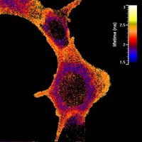
Fluorescence Lifetime Imaging (FLIM) with Femto- and Picosecond Fiber Lasers
Measuring molecule excitation lifetimes
What is FLIM?
FLIM is a microscopy technique that is based on measuring the photoluminescence (PL) decay time of either dyes used for labeling or the investigated material itself. After absorbing a photon, the fluorescent molecule stays in an excited state until it emits a photon or returns to the ground state by a non-radiative process (quenching, energy transfer). This time is characteristic for a certain material (under constant environmental conditions) and can thus be used to discriminate fluorescence signals even with strongly overlapping spectra.
The fluorescence lifetime is strongly influenced by the chemical environment in the vicinity of luminescent material. This way it is possible to get information on parameters such as local pH-value, oxygen or ion concentrations and charge transfer dynamics as well.
How does FLIM work?
FLIM is generally based on a laser scanning microscope in combination with time correlated single photon counting (TCSPC) electronics. A pulsed laser beam scans the sample pixel wise and the time between an excitation pulse and the emission of a single fluorescence photon is measured. This is repeated several times. The arrival times of the photons are registered in a histogram by the electronic. The envelope function of this histogram leads to the transient of the PL-decay. This transient can be modeled after folding the data with the instrument response function (IRF) to extract for the influence of the setup, by exponential functions which can consist of single- up to multi-exponential terms.
From this fit, the intensity averaged fluorescence lifetimes for every pixel are extracted. The result is a color-coded image of the sample. FLIM combines the 3D resolution of the confocal/two-photon microscope with a time-resolved measurement, thus giving full 4D information about the sample.
TOPTICA's offer
TOPTICA offers a broad variety of femto- and picosecond fiber lasers ideally suited for being used in FLIM microscopy, due to their short pulse durations and broad wavelength coverage.

